Intravenous Urography (IVU) and Ultrasound are two different imaging techniques used to diagnose urinary tract problems. IVU uses X-rays and contrast dye, while ultrasound relies on sound waves to create images. Both can detect kidney stones, blockages, and abnormalities, but they work in different ways.
This article outlines both tests and explains which one may be more suitable for urinary tract diagnosis.
What is Intravenous Urography (IVU)?
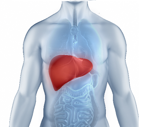
Intravenous Urography (IVU), also called Intravenous Pyelogram (IVP), is an X-ray imaging technique that is used to create images of the urinary system, including kidneys, ureters, and bladder, and assess its function.
During the test, a contrast dye is injected into a vein, which travels through the blood to the kidneys and urinary tract. The IV Urography procedure typically lasts 60 minutes, and as the dye passes through, multiple X-ray images are taken at timed intervals.
How is IVU Used for Urinary Tract Diagnosis?
Usually, the urinary tract is not clearly visible on an ordinary X-ray. It’s only when the contrast dye in the Intravenous Urography procedure is injected into the bloodstream and passes through the urinary system that the X-rays are blocked, making the structures of the kidneys, ureters, and bladder clearly visible on X-ray pictures.
An IV urogram procedure helps with the following:
- Identifies location, size, and number of kidney stones.
- Visualises abnormal growths in the kidneys or bladder.
- Shows narrowing or blockage in ureters or urinary passages.
- Assesses how efficiently kidneys filter and excrete the contrast dye.
- Reveals congenital defects or structural irregularities.
- Diagnoses the cause of blood in urine, lower-back pain, or recurrent infections.
What is Ultrasound?
Ultrasound is also a medical imaging technique that uses high-frequency sound waves to create real-time images of internal body structures. Also known as sonography, this technique uses a transducer that emits sound waves that bounce off organs and tissues, creating echoes. The echoes are captured and then converted into visual images on a screen.
An ultrasound scan is non-invasive, painless, and radiation-free, making it safe for examining organs and blood vessels, including developing foetuses, and diagnosing various medical conditions.
How does an Ultrasound help with Urinary Tract Diagnosis?
This is how a USG abdomen, or the abdominal ultrasound, which examines the urinary system, works:
- After an application of gel on the skin over the pelvic area, the transducer or ultrasound probe is placed on it, which emits sound waves that penetrate the body and bounce back from internal structures.
- High-frequency sound waves produce real-time images of the urinary tract without radiation or contrast dyes.
- The echoes are converted into detailed images on a screen, allowing doctors to instantly visualise the urinary tract.
A sonography also helps diagnose:
- Kidney stones
- Kidney abnormalities
- Bladder conditions
- Urinary obstructions
- Blood flow assessment in kidneys
- Congenital or structural abnormalities
Which Test is Better: IVU or Ultrasound?
To determine which test is better, it’s important to consider the risks or challenges each test might pose.
Although Intravenous Urography is generally considered safe, there can be some complications and challenges, such as:
- Risk of allergic reactions to the dye, which can range from mild to severe.
- Requirement for kidney function, as it’s not suitable for patients with severe kidney impairment.
- X-ray radiation exposure, which especially limits its use in pregnant women.
- A lengthy procedure time, typically lasting 30-60 minutes, with multiple timed images.
These are some of the reasons why IV urography procedure is being replaced by procedures like ultrasound, CT Intravenous Urogram, and MRI scans, which provide better imaging.
Ultrasound has no complications or side effects, and it’s considered very safe. The procedure is entirely painless, with no radiation or invasive dyes required. Ultrasound scan is also more cost-effective than IVP, provides results faster, and is preferred in children and pregnant women. However, it is frequently combined with a plain X-ray for comprehensive evaluation.
Ultimately, the better choice depends on clinical context, patient condition, and diagnostic needs.
For most urinary tract conditions, ultrasound is the preferred initial diagnostic test due to its safety profile and effectiveness. IVU may be recommended as a follow-up when functional assessment is needed or when a more detailed ureteral evaluation is required. The doctor can determine the most appropriate test based on specific symptoms, one’s medical history, and diagnostic needs.
For quick test bookings and easy access to test reports, download the Dr Lal PathLabs app now.
FAQs
- How do I prepare for an IV Urogram X-ray?
Before an IV urogram X-ray, one can be asked to fast for 4-6 hours and may need to take a mild laxative to clear the bowels. - What is the standard ultrasound price?
In India, ultrasound price typically varies, depending on the type of scan and the clinic’s location.