
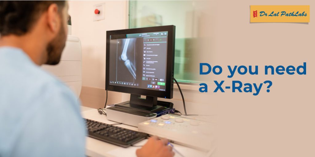
The knees are the largest joints in the human body. They are located between the two large bones of the legs, the femur (thigh bone) and tibia (shin bone). Knee joints are responsible for moving legs and body weight when standing or moving. As such, they are susceptible to injury or disorders that can cause pain or discomfort. A knee X-ray is the first step in detecting these injuries and disorders.
This article explores what a knee X-ray is, how it works, common reasons for getting one, and its expected results.
What Is a Knee X-Ray?
A knee X-ray is a procedure that uses (a form of radiation) for obtaining images of the knee structure that include the femur (thigh bone), tibia (shin bone), fibula and patella (kneecap). These X-ray images, known as radiographs, are used by doctors to examine damage to the knee bones, signs of infection, or changes in joint space.
How Does a Knee X-Ray Work?
A knee X-ray uses controlled amounts of radiation, which pass through the body and produce an image on special photographic film or digital sensors. This radiation is absorbed in different amounts by your bones and tissue because of their thickness.
The calcium in bone absorbs most of the radiation and appears white on the radiographs. Soft tissues like muscles, ligaments, tendons, and cartilage absorb less radiation and appear in varying shades of grey. Body fluids are radiolucent and thus appear in a dark tone in X-ray images.
What Are Common Signs and Reasons for a Knee X-ray?
The health of a knee joint is affected by many things. A knee X-ray is usually the first diagnostic test recommended by doctors to determine the cause of the ailment. Some of the common reasons for a knee X-ray include:
- Pain: Persistent knee pain is the most common reason for an X-ray. A knee X-ray helps in determining whether the pain is caused by arthritis, osteoporosis, or any degenerative condition that is damaging the bone or the cartilage.
- Injuries: Knee injuries from sports, falls, or accidents are another common reason for a knee X-ray. It helps in revealing fractures, cracks, dislocations, or any other signs of trauma to the knee or the bones that make up the knee joint.
- Tumours: A detailed imaging of tumours is usually obtained through an MRI or CT scan. But an X-ray can still help in identifying signs of bone tumours or abnormal growths in the knee joint. A knee X-ray is useful for identifying the size, shape, and location of the tumour.
- Joint Disorders: Knee joint disorders like osteoarthritis or rheumatoid arthritis that cause pain and affect movement in the knees are diagnosed with the help of an X-ray.
- Monitoring Progress in Medical Treatment: Knee X-rays are used for monitoring the effectiveness of treatment for bone fractures or replaced joints.
What to Expect in Knee X-ray Results?
The X-ray images of the knee joints are examined by radiologists or orthopaedists. A normal knee X-ray will vary from an abnormal one and show the following results.
- Normal Knee X-ray: A healthy knee joint X-ray would show the bones with smooth bone edges, clear gaps in bones, a perfectly centred kneecap and no signs of break, thinning, or growths on the bone.
- Abnormal Knee X-ray: An abnormal knee X-ray can show any of the following results.
- Narrowed space in the knee joint.
- Jagged, uneven knee surface showing signs of damage.
- Osteophytes or bone spurs sticking from the bone edges.
- Tiny black holes below the joint surface that indicate the presence of subchondral cysts.
- Petella (kneecap) tilted to the side or sitting too high or too low.
- Fragments of bone or cartilage floating inside the knee joint.
- Shadows of soft tissues or fluid around the knee joint that cause swelling.
Knees are the most used joints in the human body and play an essential role in walking, running, and supporting body weight. Any pain or discomfort in the knee or surrounding areas can indicate an injury or a joint disorder. It is critical to consult a doctor when experiencing knee pain and to schedule a right or left knee X-ray with Dr Lal PathLabs. Download the Dr Lal PathLabs app to schedule a test conveniently!
FAQs
- How does a normal vs abnormal knee X-ray look like?
A normal knee X-ray shows bones with smooth edges, clear gaps, a perfectly centred kneecap, and no signs of fracture, thinning, or growth on bones. An abnormal knee X-ray, on the other hand, would show narrowed space, bone fragments and shadows of soft tissues in knee joints, jagged bones, and a tilted kneecap. - How to know if knee pain is serious?
Knee pain is serious if it is swollen, shows signs of deformity, or is unable to bear any weight. A knee X-ray can help in detecting the underlying cause of the pain.