 According to the “Children in India 2025” report published by the Government of India, the annual Infant Mortality Rate in India has reduced from 44 to 25 in the last 10 years. This rapid decrease is due to better health care and diagnostic techniques to constantly monitor both fetal and maternal health during pregnancy.
Fetal echocardiography is one such diagnostic technique used during pregnancy to monitor fetal heart health. This article covers what a fetal echocardiogram procedure is, how it works and its benefits.
What is a Fetal Echo Test?
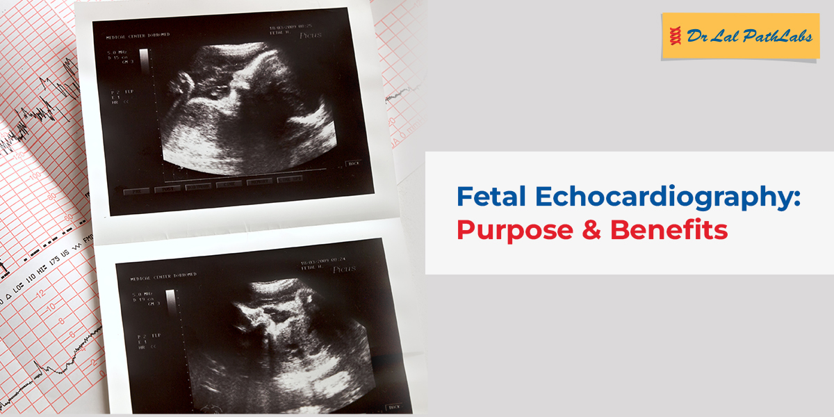
A fetal echocardiogram, or a fetal echo, means a specialised ultrasound scan that examines a baby’s heart before birth. It uses high-frequency sound waves to capture moving images of the fetal heart, including the chambers, valves, arteries, veins and blood flow pattern.
A fetal echo is different from a regular prenatal ultrasound as it focuses predominantly on the cardiovascular system. After a fetal echo test, if the baby is suspected or at risk of developing any heart conditions, the doctor can analyse the results for a more accurate diagnosis.
Why Fetal Echo is Done
A fetal echo is done to detect any congenital heart defects during pregnancy.It determines if:
- The heart’s chambers are developing at a normal rate.
- The valves of the heart are opening and closing correctly.
- There is enough blood flow through the heart, and all major blood vessels are functioning normally.
- The heartbeat has a regular rhythm.
When is a Fetal Echo Recommended?
A fetal echocardiogram is not a mandatory test for all pregnancies. It is recommended, especially when there is a higher chance of congenital heart issues. It is usually advised if:
- Either parent has a congenital heart condition.
- The fetus shows heart-related abnormalities.
- The mother has diabetes, uncontrolled hypertension or any autoimmune condition.
- The mother had certain infections during pregnancy.
- The pregnancy was achieved through assisted reproductive techniques.
- There is a prior history of genetic disorders in the family.
- The mother is taking medications known to affect fetal development.
How is a Fetal Echocardiogram Performed?
A fetal echo is a safe, painless, non-invasive procedure. The process is similar to a prenatal ultrasound but focuses specifically on the heart. During the scan:
- The expectant mother lies comfortably on an examination bed.
- A gel is applied to the abdomen to help transmit sound waves.
- A handheld probe (transducer) is moved gently across the belly.
- The sound waves create detailed, real-time images of the fetal heart on the screen.
In some cases, a transvaginal fetal echo may be performed during early pregnancy, or if the baby’s position or gestational age makes abdominal imaging challenging. The scan usually lasts 30 to 60 minutes, depending on the baby’s movements and the visibility of cardiac structures.
What are the Risks of Getting a Fetal Echocardiogram?
The fetal echo test is considered to be extremely safe for both the mother and the developing baby. It does not use radiation, and most women do not experience any discomfort beyond a slight pressure from the probe on the abdomen.
The test is also safe to be repeated multiple times in case of a high-risk pregnancy.
What are the Benefits of a Fetal Echocardiogram?
There are several benefits that a fetal echo scan offers:
- It helps with the early diagnosis of congenital heart defects. This allows medical teams to prepare for specialised newborn care.
- Leads to better pregnancy management, including decisions on delivery timing.
- Helps to monitor heart functions throughout pregnancy in high-risk cases.
- Allows parents to be emotionally prepared for heart conditions in their newborn.
Pregnant women should get a fetal echo done if they fall into a high-risk category or if their doctor suspects a possible heart-related concern during routine scans. Early screening helps monitor fetal heart development and supports timely medical decisions where required.
Always consult with a doctor to determine if a fetal echo is necessary. Download the Dr Lal PathLabs app today to book a fetal echo test!
Frequently asked questions
- What does a fetal echo test for?
A fetal echo scan checks the structure and functioning of a fetus’s heart. It checks that all chambers and valves of the heart are formed correctly and that blood flow and rhythm are normal. It detects any congenital heart defects and cardiac abnormalities before birth. - Is a fetal echo scan compulsory?
No, it is not compulsory for all pregnancies. A fetal echo is recommended only when there is a greater risk of congenital heart issues or when abnormalities are suspected during a routine ultrasound. - In which week is fetal echocardiography done?
A fetal echo is typically conducted between 18 and 24 weeks of pregnancy, when the baby’s heart is developed enough to be evaluated in detail. - Should one get a fetal echocardiogram even after a normal routine ultrasound?
Routine ultrasounds provide a general overview of fetal growth, but they cannot capture detailed heart structure and blood flow. A fetal echocardiogram offers a closer look at the heart, which makes it valuable if there are even slight risks or family history.